When fitting multifocal contact lenses, one factor of critical importance is often overlooked- the appropriate alignment of multifocal optics over a patient’s visual axis. The case study below examines the effect of altering the optical center of the multifocal lens to align more appropriately with the visual axis. Read the full case report here.
Case Study
A 45-year-old presbyopic female was fit with two sets of SpecialEyes 54 Multifocal test lenses: one that has optics positioned in the center of the lens, and one in which the optical zone is displaced 0.5mm nasally to determine its effects on visual function. Her exam findings were as follows:
Subjective Refraction:
OD -5.00 sph
OS -4.50 -.25 x 125
Add +1.50
BCVA = 20/20
Keratometry:
OD 42.98@066 / 42.14@156
OS 42.78@117 / 42.12@027
The patient’s first set of test lenses was a prism-ballasted, soft, simultaneous-design multifocal contact lens. The lenses were designed with distance optics in the center of the right lens and near optics in the center of the left lens, with the following parameters:
OD -4.75 / +2.00 distance center / 8.4 / 14.6
OS -4.25 / +2.00 near center / 8.4 / 14.6
After allowing the lenses to settle on the eye, the slit lamp examination revealed a proper contact lens fit. Next, topographies were performed over the surface of the lenses on both eyes and the optical centers were compared to the patient’s visual axis. Distance and near visual acuities were recorded both monocularly and binocularly. The patient was then asked to look at a number of targets binocularly and rate the quality of her vision on a scale from zero to 10, with 10 being the best vision and zero being the worst.
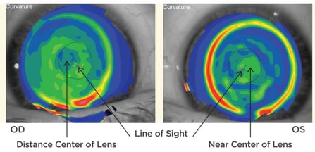
Figure 1: Right-eye and left-eye topography performed over the first set of multifocal contact lenses with optics positioned in the center of the lens. The contact lens in the right eye has distance optics in the center of the lens, while the contact lens in the left eye has near optics in the center of the lens. The large white cross represents the patient’s visual axis; the black arrows indicate the center of the optics in comparison to the patient’s visual axis. In the right-eye topography, notice the 1.12mm mismatch between the center of the distance optics and the patient’s visual axis. In the left-eye topography, notice the 0.93mm mismatch between the center of the near optics and the patient’s visual axis.
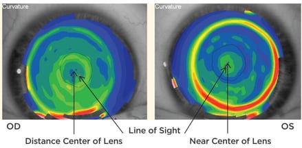
Figure 2: Right-eye and left-eye topography performed over the second set of multifocal contact lenses with 0.50mm nasally decentered optics. The contact lens in the right eye has distance optics in the center of the lens, while the contact lens in the left eye has near optics in the center of the lens. The large white cross represents the patient’s visual axis; the black arrows indicate the center of the optics in comparison to the patient’s visual axis. In the right-eye topography, notice the 0.49mm mismatch between the center of the distance optics and the patient’s visual axis. In the left-eye topography, notice the 0.30mm mismatch between the center of the near optics and the patient’s visual axis. Nasally decentering the optics by 0.50mm helped to more closely align the center of the optics with the patient’s visual axis.
The targets included:
1. The 20/30 line on the distance visual acuity chart
2. The 20/40 line on the near visual acuity chart
3. A newspaper
4. An email on the patient’s cell phone
5. The side of an eye-drop bottle
6. A phonebook
All near-sighting tasks were performed at 16 inches.
The first set of lenses was then removed and the same procedure was repeated with the second set of lenses. The second set was identical to the first except the optical centers of the lenses were displaced 0.5mm nasally on both lenses:
OD -4.75 / +2.00 distance center / 8.4 / 14.6 / 0.5mm nasally decentered / prism ballast
OS -4.25 / +2.00 near center / 8.4 / 14.6 / 0.5mm nasally decentered / prism ballast
All of the assessments performed with the first set of lenses were then performed with the second set.
Results:
First Set of Lenses:
OD 20/20-1, OS 20/25-1/+1 and OU 20/20-1 at distance
OD 20/30-2, OS 20/40-1 and OU 20/30-2 at near
Second Set of Lenses:
OD 20/20, OS 20/25-1 and OU 20/20 at distance
OD 20/40-2, OS 20/20+2 and OU 20/20+2 at near
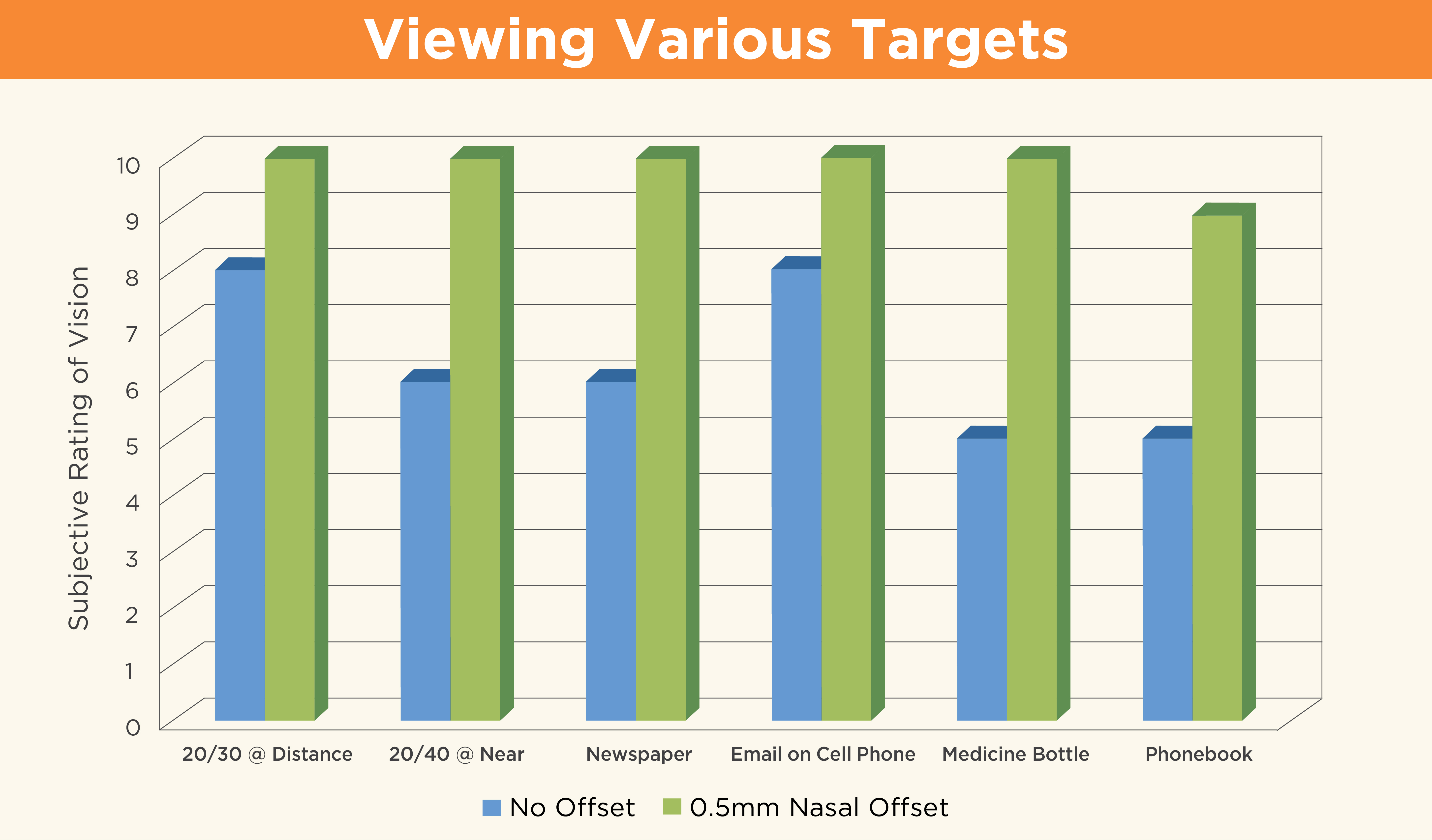
The patient’s subjective ratings while viewing the six targets are illustrated in Figure 3. These ratings tell us much more than Snellen chart visual acuity measurements alone. With the first set of lenses (no offset), the ratings for each target ranged from 5 to 8. With the second set of lenses (0.5mm nasal offset), the patient rated five targets as a 10 and the sixth target (phonebook) as a 9. Though the optical center of the lens was not directly aligned over the patient’s visual axis, it was close enough to significantly improve her subjective visual response to even some of the most critical visual targets.
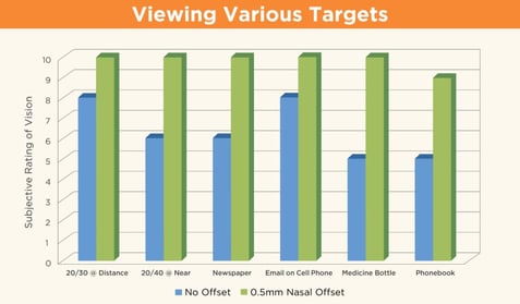
Figure 3
In conclusion, this case showed that the subject experienced enhanced visual performance when the optical center of the lens was modified to better align with her visual axis. It is believed that as additional research is conducted into the effects of optical alignment with the visual axis, there will be more findings to support this case study. In the meantime, we encourage you to check out other visual axis alignment studies listed below to learn more.
Assessing Soft Multifocal Contact Lens Centration with the Aid of Corneal Topography
How Important Is Optical Placement in Multifocal Lenses